Research
Our group focuses on the development of innovative biosensors, single cell devices, and biofuel cells based on original nano- and microchip fabrication technologies using characteristic and specific functions of nanomaterials (metal nanoparticles, carbon nanotubes etc.) and biomolecules (DNA, antibody, enzyme, cell, etc.).Interference Localized surface plasmon resonance (iLSPR) biosensor
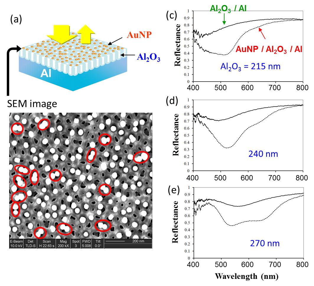 Our new sensor, that calls iLSPR sensor, was composed of metallic substrate, transparent layer, and gold nanoparticle layer. Two light waves reflected at gold nanoparticle layer and metallic substrate surface make interference, so that a fringe pattern is shown in reflection spectrum. Gold nanoparticles (AuNP) also show LSPR optical properties, the interference fringe patterns are modulated at LSPR wavelength.This effect converts broad LSPR band into a modulated fringe pattern with a high contrast. In addition, multiple reflections in the transparent layer enhance the interaction of light and gold nanoparticles. These effects are expected to make the spectral change prominent and increase the sensitivity.
References: 1)H. Minh Hiep, H. Yoshikawa, M. Saito, E. Tamiya, ACS Nano, Vol.3, 446-452 (2009), 2) H. Minh Hiep, H. Yoshikawa, E. Tamiya, Analytical Chemistry, Vol.82, 1221-1227 (2010), 3) H. Minh Hiep, H. Yoshikawa, E. Tamiya et al., Japanese Journal of Applied Physics, Vol.49, 06GM02 (2010)
Novel gold-capped nanopillars imprinted on a polymer film for highly sensitive low-cost plasmonic biosensing
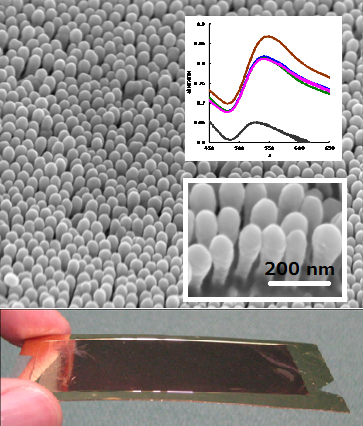 Nano-pillar structures were transferred to the resin film surface from the nano-porous almina mold by a thermal nanoimprinting process. A plasmonic substrate was fabricated by sputtering a thin layer of gold onto this nano-pillar polymer structure, and the refractive index response in a variety of media was evaluated. The biosensing capacity of this novel plasmonic substrate was verified by analysis of Human immoglobulin and achieved a minimum detection limit of 1.0 ng/mL. With the advantages of mass production with consistent reproducibility stemming from the nanoimprint fabrication process, our gold-capped polymeric pillars are ready for the transition from academic interest into commercialization systems for practical use in diagnostic applications.
Nanobio analysis using a focused laser beam
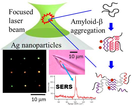 We study novel nanobio analysis based on strong molecular-photon interactions including fluorescence, Raman scattering, photochemical reactions, optical force by using a tightly focused laser beam. 1) Near infrared (NIR) optical trapping was applied to characterize the early stages of amyloid-beta aggregation in the presence of a beta-sheet intercalating dye, Congo Red (CR), as the fluorescent marker. The integration of two-photon excited fluorescence analysis with NIR optical trapping has provided a new outlook into the first two hours of amyloid-beta aggregation. 2) Plasmonic silver nanoparticle (AgNP) assemblies were fabricated on a glass substrate and in a glass micropippete by using a focused visible laser beam. This laser microfabrication of metal nanostrustures is useful for the surface enhanced Raman scattering (SERS) analysis of intercellular and intracellular signaling molecules.
Reference: A. J. Veloso, H. Yoshikawa, X. R. Cheng, E. Tamiya, K. Kerman, Analyst 136, 4164 (2011)
Rapid detection for primary screening of influenza A virus: microfluidic RT-PCR chip and electrochemical DNA sensor
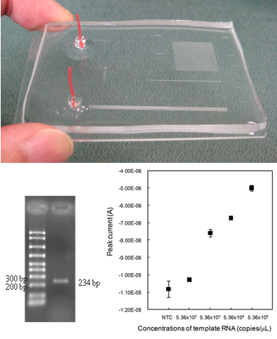 Rapid and definitive diagnosis is critical to the prevention of the spread of endemic human pathogenic viruses. Detection of variant specific genes by reverse transcription polymerase chain reaction (RTPCR) has become a routine diagnostic test for accurate subtyping of RNA viruses, such as influenza. In this research, we demonstrate the use of a continuous-flow polydimethylsiloxane (PDMS) microfluidic RT-PCR chip for rapid amplification of new influenza (AH1pdm) virus of swine-origin. The RT-PCR chip consisted of four zones: RT reaction zone, initial denaturation zone, thermal cycle zone for PCR (2-step PCR) and pressurizing-channel zone for preventing air-bubble formation. The RT-PCR was completed within 15 min which was the total flow-through time from the inlet to the outlet. This microfluidic platform for rapid RT-PCR are suitable for integration, and have the potential to be a portable system for diagnostic tests.
References: 1) Rapid detection for primary screening of influenza A virus: microfluidic RT-PCR chip and electrochemical DNA sensor, Keiichiro Yamanaka, Masato Saito, Kenji Kondoh, Mohammad Mosharraf Hossain, Ritsuko Koketsu, Tadahiro Sasaki, Naoki Nagatani, Kazuyoshi Ikuta and Eiichi Tamiya, Analyst, 2011, 136, 2064?2068, 2) An optimal design method for preventing air bubbles in high-temperature microfluidic devices, Tsuyoshi Nakayama, Ha Minh Hiep, Satoshi Furui, Yuji Yonezawa, Masato Saito, Yuzuru Takamura, Eiichi Tamiya, Anal Bioanal Chem, 396(1), 457-464, 2010
Development of micro fluidic based single cell manipulation chip
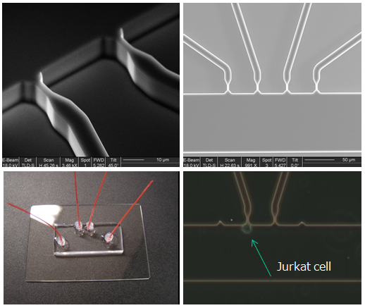 It is well known that similar cells could show different activities by separating into individual cells as revealed through further analyzing steps. Therefore isolate, analyze and recover individual cells is particularly important in the medical field such as providing safe cellular materials for regenerative medicine, or drug screening and development of human antibodies. Established isolating methods often use laser or DEP, but they both caused the bad effects due to applying heat and electricity to the cells. In this study, in order to develop microfluidic device that can recover only the isolated target cells, we designed a microchannel to trap and release a single cell only by the simple fluid controlling.
Cardiomyocyte imaging for drug testing
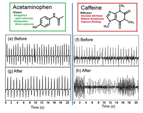 Cardiomyocyte toxicity is the most important testing for drug safely. At Tamiya laboratory, we are proposing an in-vitro quantitative imaginganalysis for cardiomyocyte dynamics. The quantitative imaging is simple and non-invasive for cardiomyocyte because the quantification was processed based on a non-invasive microscopic digital recordings. Our quantitative imaging provides the strength of the cardiomyocyte dynamics as well as the beating frequency, and is competitive to the conventional measurement. (i.e. MEA).
Reference: 1) Mohannad Mosharaf Hossain, Eiichi Shimizu, Sathuluri Ramachandra Rao,Masato Saito, Yoshinori Yamaguchi, Eiichi Tamiya, "Non-invasive characterization of Mouse Embryonic Stem cell derived Cardiomyocytes based on the intensity variation in digital beating video", Analyst, 2010,135, 1624-1630 (2010)
Raman diagnosis for the differentiation process
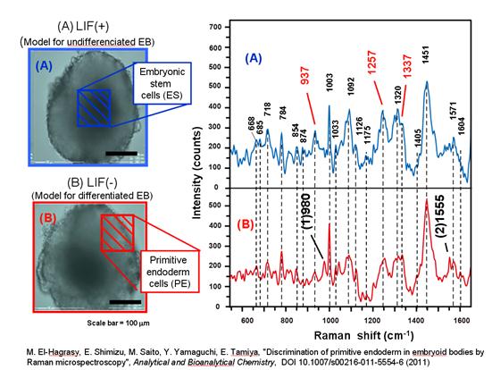 The exciting frontier for biological sensing is to monitor a stem cell differentiation. At Tamiya laboratory, we are applying Raman imaging for understanding the cardiomyocyte differentiation from mouse stem cell. The mouse stem cell is cultivated by the hanging drop (HP) to differentiation into cardiomyocytes. We are monitoring the differentiation process from the embolic body (EB) through the stem cell to the primitive endoderm (PE) by Raman spectroscopy and found the consistent Raman band among ES and PE as well as the unique Raman band for PE.
Reference: 1)Maha A. El-Hagrasy, Eiichi Shimizu, Masato Saito, Yoshinori Yamaguchi, Eiichi Tamiya, "Discrimination of primitive endoderm in embryoid bodies by Raman microspectroscopy", Analytical and Bioanalytical Chemistry, DOI 10.1007/s00216-011-5554-6 (2011)
Highly functional nanomaterials for direct biomass based fuel cells
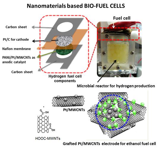 For alternative energy sources, enhancement of the power gain from biomass based-fuel cells is a desired outcome and nanomaterial intervention can offer a good leap to that end. Herein, instead of conventional inorganic transitional metal alloys and/or metal oxides, conducting polymers and functionalized multi-walled carbon nanotubes (f-MWCNTs) were used to support Platinum (Pt) as the anodic catalysts for the enhanced catalytic activity, poisonous tolerance and the utilization of Pt for cost effectiveness. Two of the most prospect biomass based-fuel cells were addressed as targets for our catalyst development: Bio-Hydrogen and Bio-Ethanol fuel cells. We also directly catalysed carbohydrates on the fuel cells. This study focused on developing the synthesis of Au nanoparticle-decorated functionalized multi-walled carbon nanotubes (Au-NPs/f-MWCNTs) for monosaccharide (bio-fuel) oxidation reactions and practical application. Another approach is synthesis of a carbon nanotube (CNT)-based catalyst that mimics the catalytic center of sugar degradation (Bio-inspired catalyst).
References: 1) Biosens. Bioelectron 25, 2509(2010) J. Mater. Chem. 21, 4068 (2011), 2) Electrochem. Commun. 13, 746 (2011), 3) Electrochim. Acta, 56, 9875 (2011), 4) Chem. Commun., 47, 7176-7178 (2011), 5) Biosens. and Bioelectron., 30, 204-210 (2011)
Printable Electrochemical Biosensors
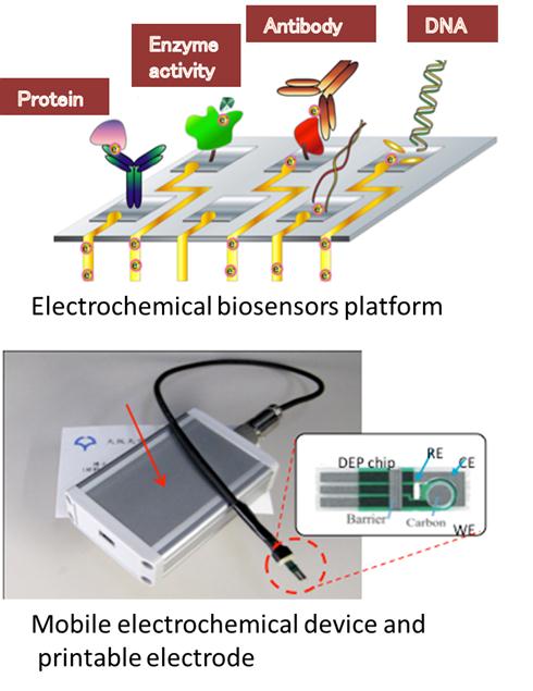 Biomarkers like genes, proteins and cellular signals can be monitored by electrochemical biosensors. Printable electrodes have advantages in mass productive and disposable applications. Our biosensors are useful for detection of pathogens like Salmonella, O-157 and Flu virus, genetic modified organism(GMO), origin of meats (Halal monitoring etc.) and so on. Gold nanoparticle-antibody can be linked with new electrochemical immunoassay. Antioxidative activity in food and amount of residual pesticides are also monitored by redox indicators and printable electrodes. Furthermore mobile an d handy electrochemical devices have been developed for biosensors aiming at POCT(point of care testing) diagnosis.
References: 1) Analyst(2011)136,2064, 2) Electroanalysis(2008)20(1)14, 3) Bioelectrochemistry (2008)74(1),118-123, 4) Food Control(2007)18(8)914, 5) Food Control(2010) 21(5) 599-605, 6) Analyst(2009) 134,966